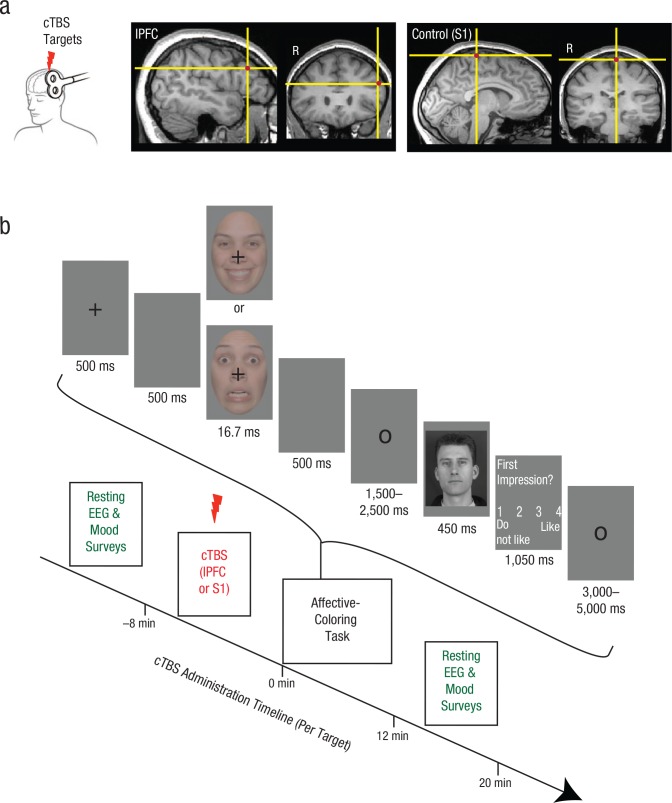
Fig. 1.
Experimental design of the transcranial magnetic stimulation (TMS) and electroencephalography (EEG) session. The brain images (a) show the lateral prefrontal cortex (lPFC) and control (primary somatosensory cortex, or S1) regions targeted during the administration of continuous theta-burst stimulation (cTBS). The target regions are overlaid on a representative subject’s T1-weighted image in native space. Each subject received TMS at both sites during the session. The timeline for each cTBS administration is shown in (b). After cTBS was administered to either left lPFC or to left S1 for 20 s, subjects completed the affective-coloring task. EEG recordings, which provided a neurophysiological index of cortical excitability, were conducted twice for each cTBS administration, before and after completion of the affective-coloring task. The trial structure for the affective-coloring task is shown in the bracketed area above the timeline. Subjects were informed that the task measured the formation of first impressions in the presence of emotional distractors. After the brief presentation of a happy or fearful face, subjects evaluated novel individuals for their likeability.

