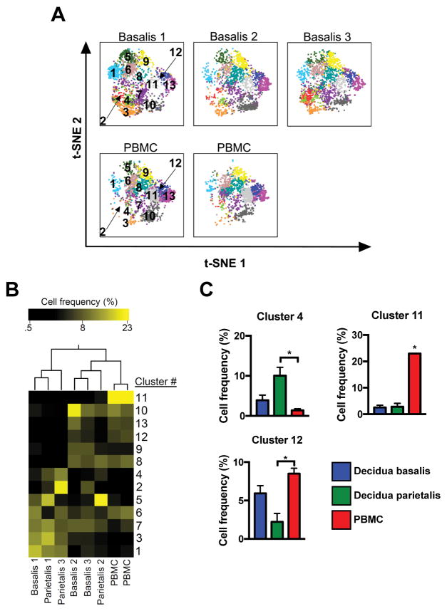
Figure 4.
Unique T cell distribution signature in term decidua. (A) Separate decidua basalis (D.B) and PBMC (P) visualized using t-SNE map generated from the merged data set. (B) Hierarchical clustering of cluster frequencies within CD3+ cells from D.B, decidua parietalis (D.P), and P. (C) Bar graphs of average cell frequencies that were determined to be statistically different. D.B (n = 3), D.P (n = 3), and P (n = 2). Significance is denoted by *p < 0.05.

