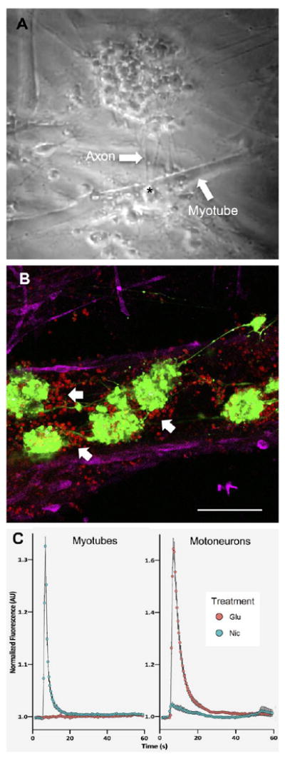
Figure 5. NMJ formation and function.

(A) A screenshot from a contraction video (See Supplementary Video S5) showed the interaction between myotubes and axons of motoneurons via NMJ after 8 days of coculture. An asterisk indicated a location of NMJ formation between axons of motoneurons and plasma membrane of myotubes. (B) Fluorescence image showed the localization of motoneurons and myotubes in coculture. Motoneurons formed a neuronal network and induced the clusterization of AChRs (showed by white arrows) in their vicinity, indicating properly developed myotubes and formation of NMJ. Red, green and magenta represent AChRs, motoneurons and actinin, respectively. Scale bar: 100 μm. (C) Normalized nicotine and glutamate-evoked calcium traces from calcium imaging on myotubes and motoneurons (See Supplementary Videos S7, S8). Nicotine could stimulate only myotubes while glutamate could trigger calcium flux only in motoneurons. Quantitative data presented as mean ± SD (n = 399 ROIs for myotubes and n = 105 ROIs for motoneurons).
