
Figure 2.
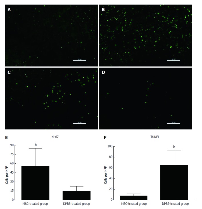
Assessment of hepatocyte apoptosis and proliferation after mesenchymal stem cell transplantation. Immunofluorescence for Ki-67 (A and B) and terminal deoxyribonucleotide transferase (TdT)-mediated deoxyuridine triphosphate nick end labeling (TUNEL) (C and D) staining in MSC-treated and DPBS-treated livers. A and C: MSC-treated group; B and D: DPBS-treated group. The numbers of Ki-67-positive and TUNEL-positive hepatocytes were observed in the DPBS- and MSC-treated groups (E and F). Bar represents the mean ± SD. (n = 5, bP < 0.001). MSC: Mesenchymal stem cell.