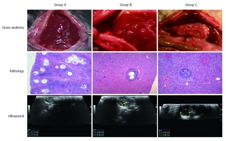
Figure 5.

Migration of the protoscolices in the portal vein and liver. The path and course of the protoscolex migration in the portal vein and liver were tracked by open examination, pathology, and ultrasound. When the human Echinococcus granulosus protoscolices were injected, they travelled from small branches of the portal vein to the liver with the blood flow, causing inflammatory cell infiltration. When the hydatid cyst formed, the infected liver presented the infection zone around the parasite lesion. The open examination showed the distribution and cyst abundance in the livers of groups A, B, and C. After 4 mo, ultrasound detected spherical, fibrous rimmed cyst with surrounding host reaction. After 6 mo, the even larger parent cyst with satellite daughter cysts within or outside the parent cyst was found.
