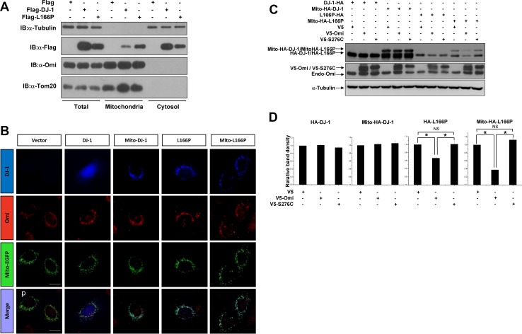
Fig. 4.
Mitochondrial targeting L166P facilitates its cleavage by Omi. A Subcellular fractionation assays of H1299 cells after transfection with Flag, Flag-DJ-1, or Flag-L166P for 36 h. B HEK 293 cells were co-transfected with plasmids expressing HA, HA-DJ-1, Mito-HA-DJ-1, HA-L166P, or Mito-HA-L166P along with Mito-EGFP as indicated. After 36 h, the cells were subjected to immunocytochemical analysis with anti-HA antibody (blue) and anti-Omi antibodies (red). Mito-EGFP was used as a mitochondrial marker (green). Scale bar, 10 μm. C Immunoblots of lysates of H1299 cells co-transfected with HA-DJ-1, Mito-HA-DJ-1, HA-L166P, or Mito-HA-L166P along with V5, V5-Omi, or V5-S276C for 36 h. D Ratios of A-DJ-1, Mito-HA-DJ-1, HA-L166P, or Mito-HA-L166P relative to α-tubulin from densitometric analysis of (B). Data from three independent experiments (mean ± SEM, *P < 0.05; ns, no statistical significance, one-way ANOVA)

