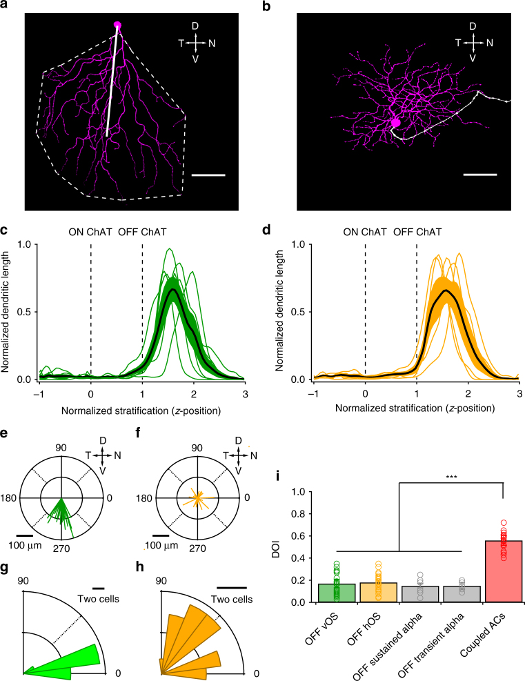
Fig. 2.
Morphology of OFF OS RGCs. a, b Dendritic morphologies of vertical OFF OS RGC (a) and horizontal OFF OS RGC (b). OFF dendrites are represented in magenta and axons are in white. Scale bar = 50 μm. Dashed line in a shows outline of dendrites. Solid line is the vector from the soma to the center of mass (COM). c, d Z-profiles of OFF vOS (c) and OFF hOS (d) dendritic arbour stratification. Thin green and orange lines indicate profiles of individual OFF vOS and OFF hOS cells, respectively. Thick black lines represent mean for OFF vOS (n = 6) and OFF hOS (n = 5). Dotted lines indicate ON and OFF starburst planes. The inner nuclear layer (INL) is located to the right and the ganglion cell layer (GCL) is located to the left. Shaded regions indicate SEM across cells. e, f Polar plots of COM vectors of OFF vOS (n = 24) (e) and OFF hOS (n = 22) (f) RGCs. Scale bar = 50 μm. g, h Rose plots of absolute differences between OS angle (θ OS) and COM vector angle (θ COM vector) of ON vOS RGCs (n = 24) (g), and OFF hOS RGCs (n = 22) (h). i Dendritic orientation index (DOI) of OFF OS RGCs (n = 26 OFF vOS and 23 OFF hOS), OFF sustained alpha (n = 6), OFF transient alpha (n = 5) and amacrine cells coupled to OFF OS RGCs (n = 19). ***p < 0.001

