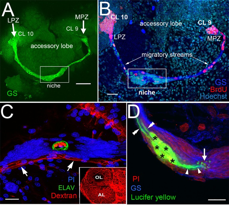
Figure 2.
The proliferative system maintaining adult neurogenesis in the crayfish (Procambarus clarkii) brain. (A) The lateral (LPZ) and medial (MPZ) proliferation zones are contacted by the processes of a population of cells immunoreactive to GS (green) whose somata are located in a neurogenic niche (white box) on the ventral surface of the brain. (B) Left side of the brain of P. clarkii labeled immunocytochemically for the S-phase marker BrdU (red). Labeled cells are found in the LPZ contiguous with Cluster 10 (CL 10) and in the MPZ near Cluster 9 (CL 9). The two zones are linked by a chain of labeled cells in a migratory stream that originates in the boxed region labeled “niche.” Labeling for glutamine synthetase (blue), BrdU (red), and Hoechst (cyan) is shown. (C) The vascular connection to the cavity in the center of the niche was demonstrated by injecting dextran tetramethylrhodamine dye into the cerebral artery. The cavity, outlined by its reactivity to an antibody to ELAV (green), contains the dextran dye (red), which is also contained within a larger blood vessel that lies beneath the niche (arrows). PI (blue) labeling of the niche cell nuclei is also shown. Inset: dextran-filled vasculature in the olfactory (OL) and accessory (AL) lobes on the left side of the brain. (D) Niche cells (green), labeled by intracellular injection of Lucifer yellow, have short processes (arrowheads) projecting to the vascular cavity (arrow) and longer fibers (double arrowheads) that fasciculate to form the tracts projecting to the LPZ and MPZ, along which the daughters of the niche cells (2nd-generation neural precursors) migrate. Glutamine synthetase (GS), blue; propidium iodide (PI), red. Scale bars: (A), 100 μm; (B), 30 μm; (C,D), 20 μm (A, C and D from Sullivan et al., 2007a).

