Figure 6.
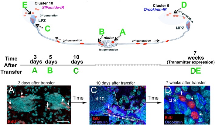
EdU+ cells were observed in the niche, streams, and in Clusters 9 and 10 of recipient crayfish after adoptive transfer of EdU+ hemocytes from donor animals. The schematic diagrams illustrate (top) the locations of the EdU+ cells that were observed following adoptive transfers, and (middle) the experimental timeline for each sample (time after transfer, A–E), and (bottom) images from P. clarkii (A,D) and P. leniusculus (C). All samples in these images (A,C,D) were labeled with the nuclear marker Hoechst 33342 (cyan). (A) Three days after adoptive transfer of hemocytes, EdU+ cells (red) were observed in the niche. (C) Ten days after hemocyte transfer, cells were observed in the distal ends of the streams, near Clusters 9 and 10 (arrow). Immunoreactivity for tyrosinated tubulin (blue) highlights the migratory stream, which is also outlined in white. (D) Seven weeks after adoptive transfer of hemocytes from donor to recipient crayfish, EdU+ cells (red) were observed in cell Clusters 9 and 10. Some of these cells in Cluster 9 express orcokinin, a peptide transmitter used by many Cluster 9 cells. Examples of cell labeling at B (5 days, in the niche) and at E (7 weeks, in Cluster 10) are not shown (but see Benton et al., 2014). Scale bars: (A), 40 μm; (C), 10 μm; (D), 10 μm. Adapted from Benton et al. (2014).

