Figure 1.
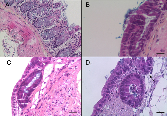
Histological validation of the murine colonic explant culture system (A) Hematoxylin eosin safranin (HES) staining of a normal colon section of an uninfected SCID mouse. Scale bar, 40 µm. (B) HES staining of a colonic explant section after 14 days of culture showing good tissue preservation with a characteristic collagen fiber and crypt-like structures. Scale bar, 20 µm. (C,D) HES staining of a colonic explant section after 21 and 30 days of culture, respectively. Tissue preservation was confirmed with a presence of high prismatic epithelium, basal position of nucleus, a well-organized basal lamina and the presence of crypt-like structures (black arrow (D)). Presence of large cyst-like formations (white arrow (C)). Scale bars, 20 µm.

