Figure 3.
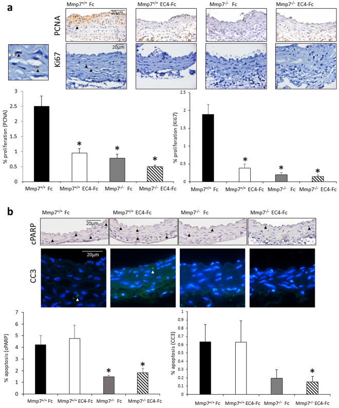
Combined Mmp-7 deletion and EC4-Fc over-expression resulted in reduced vessel wall proliferation and apoptosis in Ang-II induced abdominal aortic aneurysms in Apoe−/− mice. (a) Representative images of proliferation markers: PCNA and Ki67 immunohistochemistry in the mouse abdominal aortae. Positive cells are brown, negative nuclei are blue. Arrows indicate some positive cells. Scale bar indicates 20 µm. Small panel (left-handside) shows high power of Ki67 immunohistochemistry with two positive (brown cells). Analysis of immunohistochemistry is shown in the graphs. Proliferation was measured by counting the proportion of positive cells. * indicates a significant difference from Mmp-7+/+ Fc. (mean + SEM, n = 17–19, p < 0.05, ANOVA, Student Newman Keuls Post test). (b) Representative images of apoptosis markers: cleaved PARP (cPARP) and cleaved caspase-3 (CC3) immunohistochemistry in the mouse abdominal aortae. For cPARP: positive cells are brown, negative nuclei are blue. For CC3: positive cells are green, negative nuclei are blue. Arrows indicate some positive cells. Scale bar indicates 20 µm. Analysis of immunohistochemistry is shown in the graphs. Apoptosis was measured by counting the proportion of positive cells. * indicates a significant difference from Mmp-7+/+ Fc (mean + SEM, n = 17–19, p < 0.05, Student Newman Keuls Post test for cPARP, unpaired t test for CC3).

