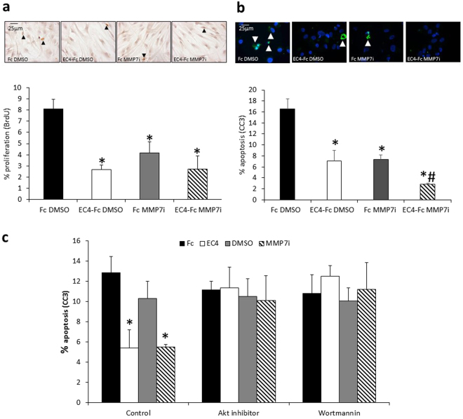
Figure 4.
EC4-Fc and MMP-7 inhibitor reduced proliferation and apoptosis of human aortic SMCs. (a) Human aortic VSMCs were treated with 20ng/mL PDGF + 20ng/mL bFGF to stimulate proliferation for 24 hours with BrdU, and 100pM Fc or EC4-Fc and 1% DMSO or 0.1 µM MMP-7 inhibitor, then BrdU incorporation quantified. * indicates a significant difference from Fc DMSO (mean + SEM, n = 4, p < 0.05, ANOVA, Student Newman Keuls Post test). Representative images of BrdU immunocytochemistry. Arrow heads indicate some positive cells, in brown. Nuclei are shown in blue. Scale bar indicates 25 µm. (b) Human aortic smooth muscle cells were treated with 200ng/mL Fas-L for 24 hours to induce apoptosis, in the presence of 100pM Fc or EC4-Fc and 1% DMSO or 0.1 µM MMP-7 inhibitor. Apoptosis was measured by cleaved caspase-3 (CC3) and the percentage of positive cells was counted. * indicates a significant difference from DMSO Fc. # indicates a significant difference from all other conditions (mean + SEM, n = 4, p < 0.05, ANOVA, Student Newman Keuls Post test). Representative images of cleaved caspase-3 immunocytochemistry. Arrowheads indicate positive cells in green. Nuclei are shown in blue. Scale bar indicates 25 µm. (c) Human aortic smooth muscle cells were treated with 200ng/mL Fas-L for 24 hours to induce apoptosis, in the presence or absence of 100pM Fc or EC4-Fc and 1% DMSO or 0.1 µM MMP-7 inhibitor as well as 10 µM Akt inhbitor or 20 nM PI3-kinase inhibitor (Wortmannin). Apoptosis was measured by cleaved caspase-3 (CC3) and the percentage of positive cells was counted. * indicates a significant difference from Control Fc (mean + SEM, n = 4, p < 0.05, ANOVA, Student Newman Keuls Post test).

