Fig. 1.

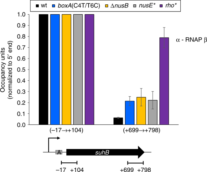
Transcription termination within suhB is dependent on Rho and Nus factors. RNAP (β) enrichment at suhB 5′ and 3′ regions was measured using ChIP-qPCR in wild-type MG1655, boxA(C4T/T6C), ΔnusB, nusE(A12E) or rho(R66S) mutant strains. Values are normalized to signal at the 5′ end of suhB. x-axis labels indicate qPCR amplicon position relative to suhB. Error bars represent ±1 standard deviation from the mean (n = 3). A schematic depicting the suhB gene, the transcription start site (bent arrow) and boxA (grey rectangle) is shown below the graph. Horizontal black lines indicate the position of PCR amplicons
