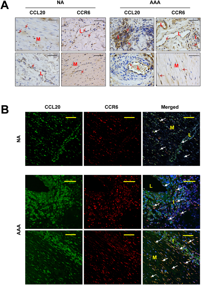
Figure 5.
(A) Representative immunohistochemistry images of CCL20 and its receptor (CCR6) in normal aorta (NA) and in AAA samples. Arrows show some immunostained cells. (B) Representative immunofluorescent double staining for CCL20 and CCR6 in normal aorta (NA) and in AAA samples. Arrows show double immunostained cells. Bars are 50 µm; L, indicates the light of microvessels; I, indicates leukocyte infiltration areas; and M, indicates media layer.

