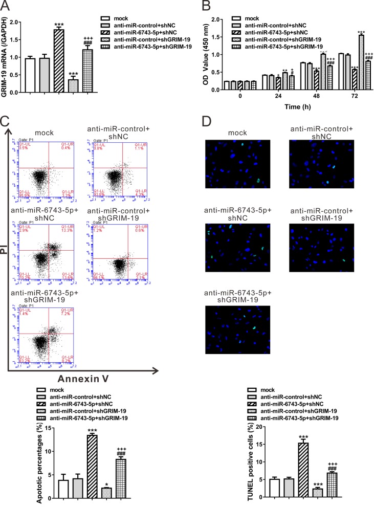
Figure 3. Opposite effects of miR-6743-5p and GRIM-19 on glioma cell proliferation and apoptosis.
U251 cells were transfected with anti-miR-control/anti-miR-6743-5p, and infected with shGRIM-19 or shNC lentivirus as indicated. Cells without any treatment were set as a negative control (Mock). (A) The mRNA levels of GRIM-19 were assessed at 48 h after treatment. (B) The proliferation curves of U251 cells at 0, 24, 48, and 72 h after treatment as determined by CCK-8 assay. (C, D) Cell apoptosis was determined by Annexin V/PI staining and flow cytometry analysis (C) and TUNEL assay (D) at 48 h after treatment. The representative images and the quantitative analysis are shown. The lower right quadrant (Annexin V+/PI–) represents the apoptotic cells. *P<0.05, **P<0.01, and ***P<0.001 compared with Mock and anti-miR-control + shNC; #P<0.05 and ###P<0.001 compared with anti-miR-6743-5p + shNC; +P<0.05 and +++P<0.001 compared with anti-miR-control + shGRIM-19.

