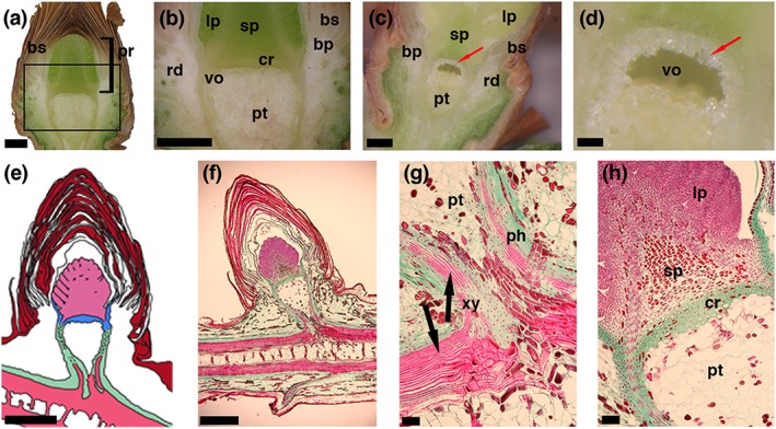
Figure 1.

Images and a schematic drawing of longitudinal sections through vegetative buds of Picea abies. (a,b) Digital images of a longitudinal section through an unfrozen (April 2016) and (c,d altered from [Neuner & Hacker, 2012]) frozen bud (June 2008). Red arrows (c,d) indicate location of ice crystals accumulated by translocated ice formation (extraorgan freezing). (e) Schematic drawing of a longitudinal section through the bud, where the ice barrier tissue (blue) formed by the crown and the inner bark tissue (in 3D a bowl‐shaped structure) encapsulates the primordium (light pink). (f,g,h) Microscopic images of a longitudinal semithin section of a vegetative bud of Picea abies before a seasonal freezing event after triple staining with safranin, orange G, and fast green. (g) Enlargement of the lower box shown in F. (h) Enlargement of the upper box in F. The phloem is colored green, the xylem pink, and the bud scales are colored red‐brown. Horizontal bars indicate 1 mm in a, b, c, e and f, 0.25 mm in d, and 0.1 mm in g and h. Abbreviations: bs = densely packed bud scales; pr = primordium; sp = shoot primordium; lp = leaf primordia; pt = pith tissue; rd = resin ducts; bp = basal part of the bud scales, cr = crown; vo = void between the crown and the pith tissue; ph = phloem; xy = xylem
