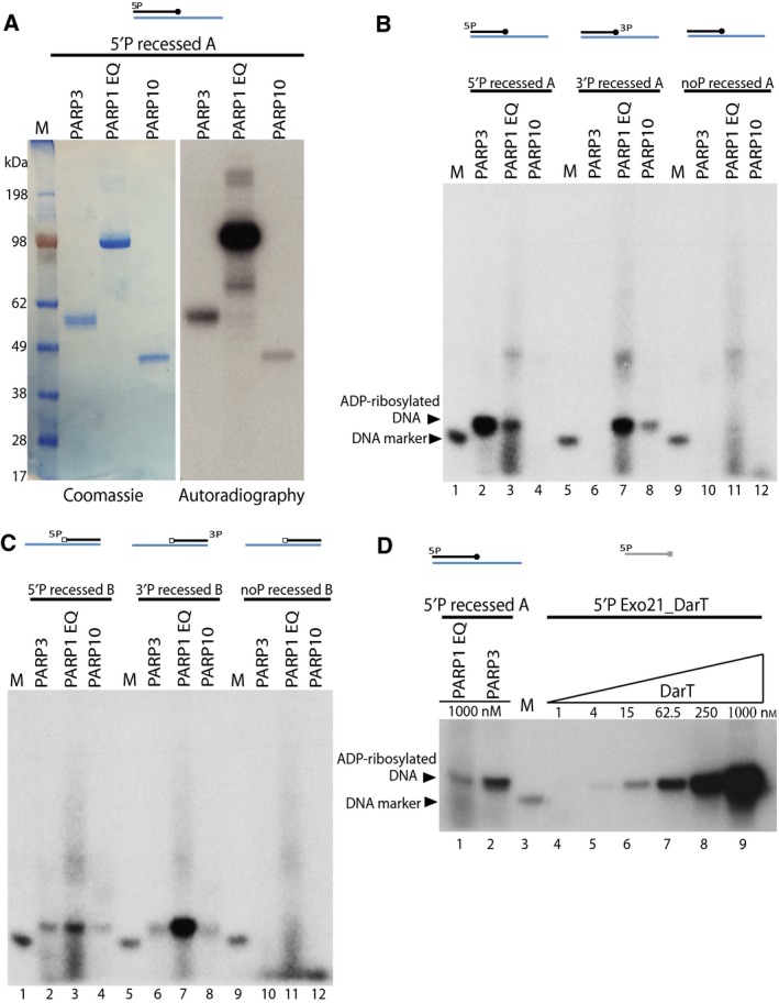
Figure 2.

Mono ADP‐ribosylation of DNA ends by PARPs. (A) Auto‐ADP‐ribosylation of PARP3, PARP1 E998Q and PARP10 – left panel shows the coomassie staining and right panel shows the autoradiography of the SDS/PAGE gel. (B) Mono‐ADP‐ribosylation of phosphorylated or nonphosphorylated recessed A DNA substrate catalysed by PARP3, PARP1 E998Q or PARP10 as seen from autoradiograph of a denaturing urea PAGE gel (C) ADP‐ribosylation of phosphorylated or nonphosphorylated recessed B DNA substrate by PARP3, PARP1 E998Q or PARP10. (D) Comparison of DNA ADP‐ribosylation activity of PARP3 and PARP1 E998Q on 5′P recessed A substrate against DNA ADP‐ribosylation of DarT on single stranded Exo21_DarT oligo.
