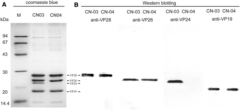
Figure 2.

SDS-PAGE and Western blot analysis of purified WSSV virions. A Equal amounts of purified WSSV-CN03 and WSSVCN04 virions were lysed, separated on 12% SDS-PAGE gel, and stained with coomassie brilliant blue. B For Western blotting, proteins separated on SDS-PAGE gels were transferred to PVDF membranes. The membranes were probed with indicated primary antibodies, and then incubated with alkaline phosphatase-conjugated goat anti-mouse IgG. The signals were detected using the NBT/BCIP substrate.
