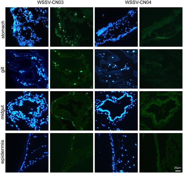
Figure 5.

Immunofluorescence analysis of WSSV-CN03 and -CN04 infection in juvenile Litopenaeus vannamei . L. vannamei were randomly divided into three groups. Each shrimp was fed with 2 mg WSSV-CN03-infected tissue or WSSV-CN04-infected tissue. The shrimp in the control group were fed with WSSV-free tissue. At 24 hpi, 5 shrimp were randomly selected from each group for cryosectioning. The slices were probed with anti-VP28 antibody, and the nucleus was stained with DAPI. Bar 20 μm. The experiment was repeated three times and typical results of one experiment are shown.
