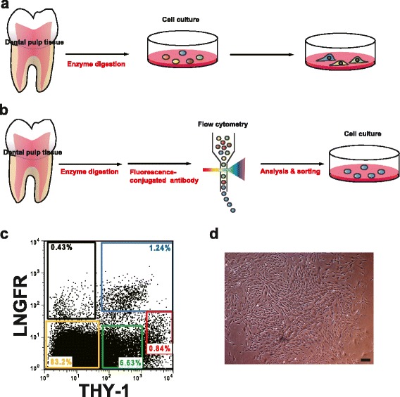
Fig. 1.

a Traditional isolation of dental pulp stem/progenitor cells (DPSCs) by adherent culture on dishes. b Prospective isolation of DPSCs by flow cytometric identification of cell surface markers. c Representative fluorescence-activated cell sorting profiles of dental pulp cells. d A representative phase-contrast micrograph of plastic-adherent colony-forming LNGFRLow+THY-1High+ cells with fibroblast morphologies. Scale bars = 100 μm
