
Fig. 6.
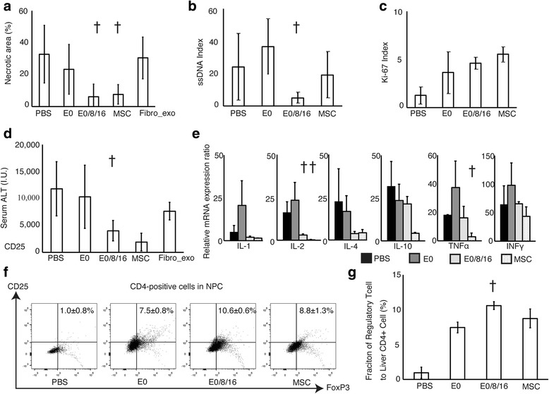
In vivo effect of exosomes derived from MSC. mRNA expression of pro- and anti-inflammatory cytokines and flow cytometric analysis of Treg among NPCs. a–c Percentage of necrotic areas in the injured liver evaluated with hematoxylin-eosin staining (a), ssDNA index (b), and Ki-67 index (c) of mice received intravenous injections of PBS only (PBS), MSC-derived exosomes once (E0), MSC-derived exosomes three times (E0/8/16), or MSCs (MSC). d Plasma ALT levels. e mRNA expression ratios of pro- and anti-inflammatory cytokines. f, g Dot plots (f) and percentage (g) of Treg to CD4-positive percentage of Treg to CD4-positive cells among NPCs. † p < 0.05 vs. the group that received a single injection of exosomes