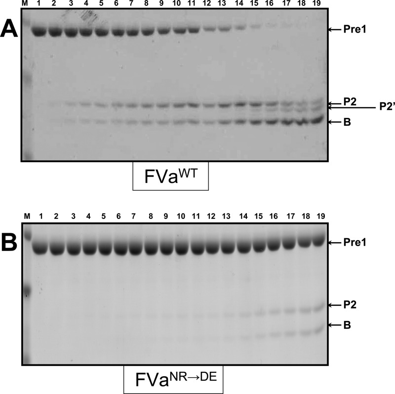
Figure 9.
Gel electrophoresis analyses for cleavage of Prethrombin-1. Prethrombin-1 was incubated in different mixtures with PCPS vesicles and rhfVa as described previously in detail.20 The reaction and the samples were further treated, scanned, and quantified as detailed in the Experimental Section. Panel A, control, rhfVaWT; panel B, rhfVaNR→DE. M represents the lane with the molecular weight markers (from top to bottom): Mr 50 000, Mr 36 000, Mr 22 000. Lanes 1–19 represent samples from the reaction mixture before and after the addition of fXa as previously described.20 The hPro-derived fragments are shown as detailed in the legend to Figure 3. The fragment denoted as P2′ depicts Pre2 cleaved at Arg284. For easy reading of the article, the rhfVa species used for the reconstitution of prothrombinase is also shown under each panel.

