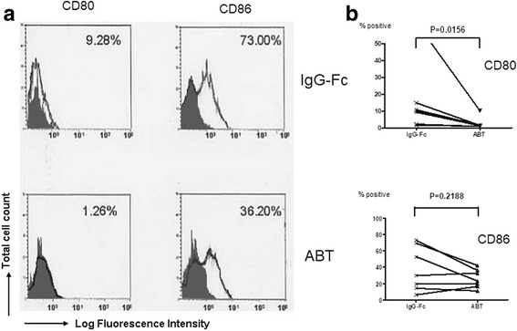
Fig. 3.

Effects of abatacept on the expression of CD80 and CD86 on SEB-stimulated monocytes. Highly purified monocytes (1 × 10⁶/well) from seven healthy individuals were cultured in the presence of SEB (100 pg/ml) in each well of 24-well flat-bottomed microtiter plates with abatacept (100 μg/ml) or control IgG-Fc (100 μg/ml) for 48 h, after which the cells were stained with FITC-conjugated anti-CD80, anti-CD86, or control IgG1, followed by counterstaining with PE-conjugated annexin V. The cells were then analyzed by flow cytometry. a Representative histograms of the staining of various molecules on annexin V-negative monocytes. The percentages of positive cells for specific mAb staining are indicated. Stainings with isotype-matched control mAb (control IgG1) are indicated by shade. b Percentages of positive cells for each specific mAb staining of monocytes from seven independent experiments are summarized. Statistical significance was evaluated by Wilcoxon’s signed-rank test
