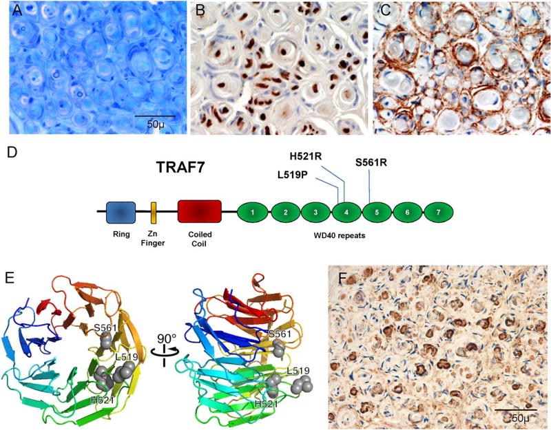
FIGURE 1.
Typical histology and gene discovery in intraneural perineurioma. (A–C) Illustrative case (A) epoxy section at similar level and magnification as shown in paraffin preparations with pseudo–onion bulb formations around occasionally seen thin myelinated fibers with (B) Schwann cell preparation (S-100) demonstrating sparse reactivity of the Schwann cells at the center and (C) prominent epithelial membrane antigen (EMA)-reactive staining of the leaflets of the pseudo–onion bulbs. (D) Functional domains and location of recurrent TRAF7 mutations. The WD40 domain is the region with identified mutations in meningiomas. (E) Rainbow cartoon representation of a model of TRAF7 WD40 domain structure. The 3 residues mutated in intraneural perineuriomas are highlighted as gray spheres and cluster together on the protein predicted to interfere with TRAF7 interaction with other proteins and/or to cause instability of the WD40 domain. (F) TRAF7 immunoreactivity in perineurioma occurs in the same areas that reacted to EMA.

