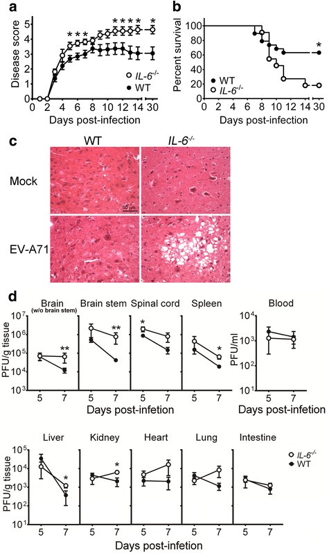
Fig. 2.

Absence of IL-6 increases the disease severity, mortality, tissue damage, and tissue viral loads of EV-A71-infected mice. The disease severity (a) and survival (b) of infected wild-type mice (black symbols or WT; n = 19) and infected IL-6 −/− mice (white symbols; n = 11) were monitored for 30 days. (c) Spinal cords of mock-infected or infected wild-type and IL-6 −/− mice were harvested 10 days after infection, sectioned, and stained with hematoxylin-eosin. The motor horn of lumbar region is shown. Data are representative of at least 3 samples from two independent experiments. (d) Viral titers in the indicated tissues of wild-type mice (WT; black circles) and IL-6 −/− mice (white circles) collected on 5 or 7 days after infection are shown. Data show means ± SEM (error bars) in panel a and 6–7 samples per data point in panel d. *P < 0.05 and **P < 0.01 compared between wild-type and IL-6 −/− mouse groups at the indicated time
