
Figure 2.
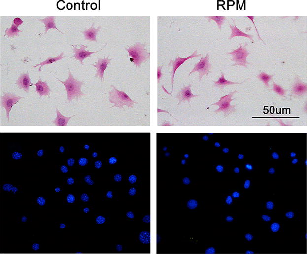
Cell morphology of MLO-Y4 cells was not altered after 2 h of RPM treatment. Cells were stained with hematoxylin and eosin (upper panels) or Hoechst dye (5 μg/ml) (lower panels) and observed under phase or fluorescence microscope. Bar, 50 μm.