
Figure 4.
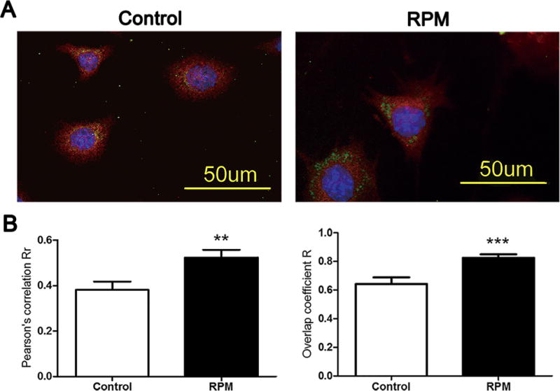
Cx43 was retained in the Golgi bodies after 2 h of RPM. MLO-Y4 cells were double labeled with antibodies against Cx43 (red, 1:300) and 58 K Golgi marker (Green, 1:50) antibodies and, followed by rhodamine and FITC-conjugated secondary antibodies, respectively (A). The degree of colocalization of Cx43 and 58 K was analyzed using the Pearson’s correlation coefficient (Rr) (left panel) and Overlap coefficient (R) (right panel) using the Image J software (B). **p < 0.01; ***p < 0.001.