
Figure 3.
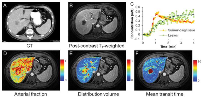
Free-breathing liver perfusion imaging using through-time spiral generalized auto-calibrating partially parallel acquisition acceleration (GRAPPA). Representative images acquired from a patient with metastatic lung adenocarcinoma. One hundred volumes were acquired continuously over a period of approximately 4 minutes with a spatial resolution of 1.9×1.9×3 mm3. (A) Computed tomography image; (B) Representative free-breathing liver DCE image acquired using through-time spiral GRAPPA; (C) Single-voxel GBCA concentration curves from both lesion and surrounding tissue. Fitted data using a dual-input single-compartment model were also plotted. Corresponding perfusion maps including arterial fraction (%), distribution volume (%) and mean transit time (sec) are shown in D–F (Courtesy of Drs. Yong Chen and Vikas Gulani).