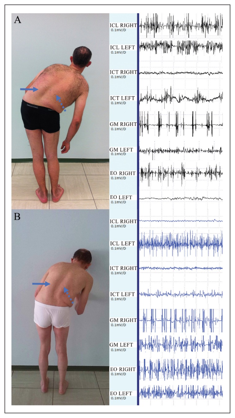
Figure 1.
A: case 5 with 50° of PS, pattern I (subtype II). He shows left (arrow) and right (dotted arrow) ICL hypertrophy. The intensity of contralateral ICL activation is reduced when compared with the right side, which indicates a possible impairment of the paraspinal compensatory mechanism. B: case 4 with 42° of PS, pattern II. He shows left (arrow) and right (dotted arrow) ICL hypertrophy. The intensity of contralateral ICL activation is increased when compared with the right side, which indicates possible integrity of the paraspinal compensatory mechanism. In both these PS patients there is hyperactivity of EO, right side.

