
Figure 7. Representative traces of increases in [Ca2+]i produced by NMDA in DRG neurons.
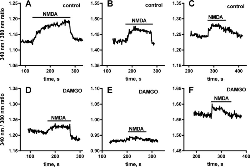
Cultured DRG neurons were loaded for 1 h with 5 μM Fura-2 AM and then incubated for 15 min with 20 ng/ml BDNF alone (A–C) or with 1 μM DAMGO (D–F). [Ca2+]i was measured with a fluorescence microscope while the cells were superfused with medium. Addition of 250 μM NMDA + 10 μM glycine (“NMDA”) produced increases in [Ca2+]i. Preincubation with DAMGO resulted in smaller increases in [Ca2+]i induced by NMDA + glycine.