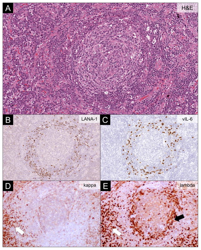
Figure 3.
Lymph Node Findings in KSHV-associated Multicentric Castleman Disease. (A) Hematoxylin and eosin (H&E) stain showing typical features of KSHV-associated multicentric Castleman disease. The involved lymph nodes have a regressed germinal center surrounded by layered mantle cells, vascular proliferation and hyalinization, and plasmacytosis in the interfollicular regions. (B) LANA-1 immunohistochemistry highlighting KSHV-infected plasmablasts residing in the mantle cell layers. (C) vIL-6 immunohistochemistry showing vIL-6 in a proportion of KSHV-infected plasmablasts. (D-E) Kappa and lambda light chain immunohistochemistry showing restricted lambda expression in the KSHV-infected plasmablasts (black arrow), while the interfollicular plasma cells are polytypic (white arrow).

