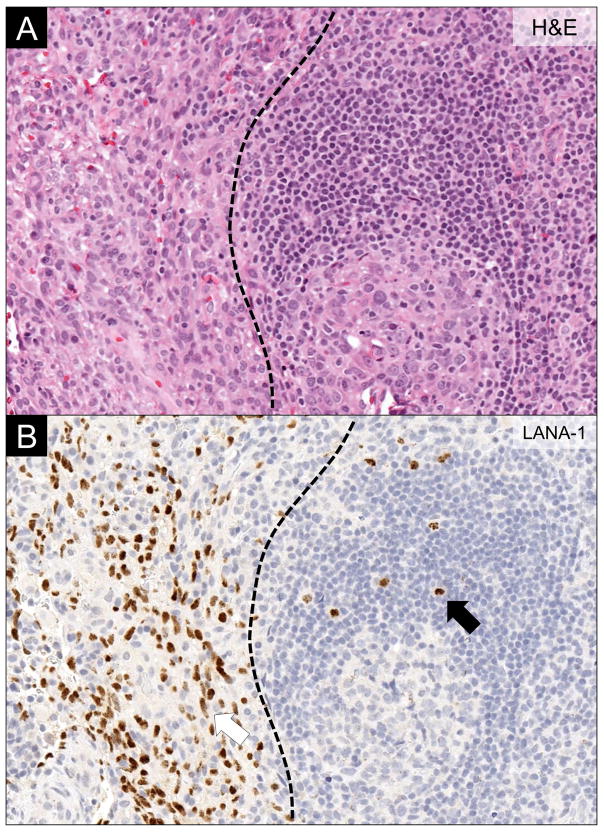
Figure 4.
Concominant Lymph Node Kaposi Sarcoma and KSHV-associated Multicentric Castleman Disease. (A) H&E stain demonstrating concomitant Kaposi sarcoma (left of the dotted line) and multicentric Castleman disease (MCD) (right of the dotted line). (B) LANA-1 immunohistochemistry highlighting the KSHV-infected cells. Note the different cytomorphology of Kaposi sarcoma cells (red arrow) and the plasmablasts in MCD (black arrow).

