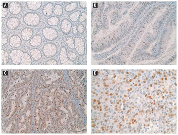
Figure 2.
Histopathologic Images of Survivin Staining. (A) Normal Colonic Mucosa Showing Undetectable Levels of Survivin Protein. (B) Tubulovillous Adenoma with Several Scattered Survivin-Positive Epithelial Cells. (C) A Well-Moderately Differentiated Adenocarcinoma Exhibiting Strong Survivin Protein Expression in Many of the Tumor Cells. (D) A Poorly Differentiated Adenocarcinoma shows Strong Survivin Staining in Most of the Tumor Cells

