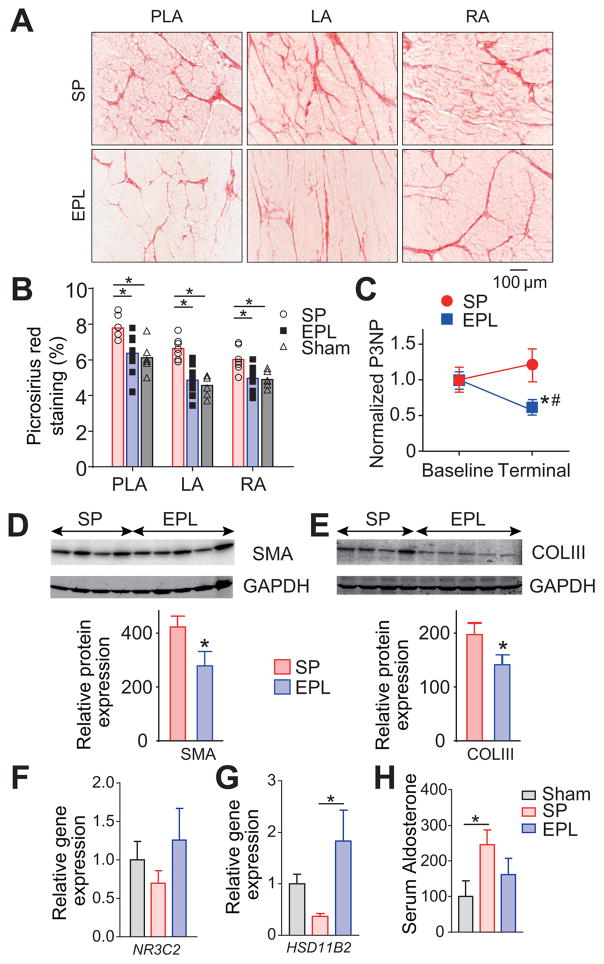
Figure 2. EPL reduces fibrosis development during AF progression.
(A) Representative picrosirius red staining of PLA, LA and RA. (B) Interstitial fibrosis for sham operated (N=6), SP-treated (N=7) and EPL-treated groups (N=10). Ten pictures per slide were randomly selected and analyzed. *P<0.05, **P<0.01. (C) EPL mitigates the increase of serum Procollagen III N-Terminal Propeptide (P3NP) during AF progression. *P<0.05 vs. Baseline, #P<0.05 vs. EPL. (D, E) Western blots for smooth muscle actin (SMA) and collagen III (COLIII) in atrial homogenates relative to GAPDH from SP-treated (N=4) and EPL-treated groups (N=5). (F,G) Mineralocorticoid receptor (NR3C2) and 11b-HSD2 (HSD11B2) gene expression in the LA. (H) serum aldosterone in sham operated, AF sheep treated with SP and AF sheep treated with EPL. For F–H, Sham (N=7), SP-treated (N=8) and EPL-treated groups (N=5). N=number of animal. *P<0.05. (B, F–H) One-way ANOVA followed by post hoc Bonferroni’s test. (C) Two-way ANOVA followed by post hoc Bonferroni’s test.

