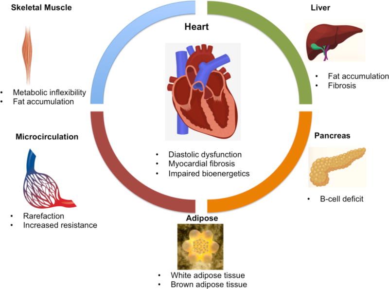
Figure.

The cardiac consequences of cardiometabolic disease ensue from functional and structural alterations in multiple organ systems, including skeletal muscle, liver, the pancreas and microcirculation. Primary imaging targets for non-invasive investigation of CMD are listed. Multiple imaging modalities are used in the evaluation of organ systems involved in CMD. Skeletal muscle investigation typically involves multinuclear MR spectroscopy and MR imaging. Liver is non-invasively interrogated using CT, elastography (both MR and US) and PET for fat quantification, fibrosis quantification and glucose utilization, respectively. Pancreatic beta cell deficit is measured in humans using novel SPECT and PET techniques and in preclinical models with magnetic nanoparticles and MR imaging techniques. Adipose tissue can be quantified using MRI, CT, US and dual-energy x-ray absorptiometry. Brown adipose tissue can be quantified using FDG-PET techniques. Microcirculatory rarefaction and resistance can be evaluated with optical imaging techniques and PET. Finally, the heart in CMD can be investigated using US, MRI and multinuclear MR spectroscopy.
