Figure 2.

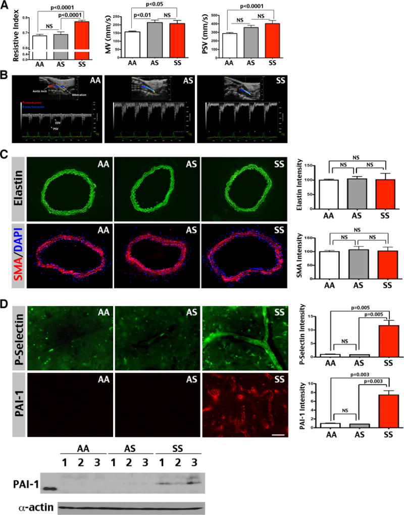
Six-month-old SS mice show increased resistive index (RI) and flow velocity (MV and PSV), but no sign for proliferative-obstructive vasculopathy in the carotid artery. (A) Six-month-old SS mice (n=9) showed a greater RI (0.77±0.0; AA: 0.68±0.01; AS: 0.69±0.02), MV (208.3±18.94 mm/sec; AA: 157±5.41; AS: 214.3±15.46), and PSV (403±35.69 mm/sec; AA: 286.1±12.36; AS: 356.4±25.99) in the carotid artery compared than AA (n=7) and AS (n=8) mice. Shown are mean ± SEM. The p-values were determined by one-way ANOVA and post hoc Newman-Keuls test. NS: p>0.05. (B) Typical Doppler pulse-waveforms in the carotid artery of AA, AS, and SS mice. (C) The carotid arteries of AA, AS, and SS mice showed comparable intensity for the internal elastic membrane (green) and anti-smooth muscle actin labeling (SMA, red), counter-stained by DAPI (blue, n=3 for each genotype). The immunostaining intensity was quantified and compared by t-test. Note that SS mice showed no evidence of duplication-interruption of the internal elastic lamina or hypertrophy of peri-vascular smooth muscle that was detected in SCA patients with overt stroke.4,5 (D) Immunofluorescence labeling showed ectopic P-Selectin (a marker for endothelial activation) and Plasminogen Activator Inhibitor-1 (PAI-1) expression in the cerebral blood vessel wall of SS mice, but not in AA or AS mice (n=3 for each genotype). The intensity of immunostaining was quantified for comparison by t-test. Likewise, immunoblotting showed consistent anti-PAI-1 signals in SS, but not AA or AS mouse brains. Shown in immunoblot are 3 mice for each genotype. Scale bar: 40 μm in C, 50 μm in D.
