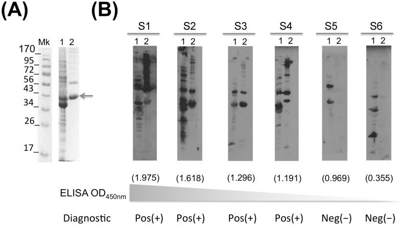
Fig 3. Immunoblot confirmation assay.
Borrelia turicatae whole cell lysates were separated in 12% SDS-PAGE gels (line 1) together with rGlpQ (line 2) and stained with coomassie briliant blue (A). Western blots were run to confirm the ELISA sero-reactivity (B). S1 through S6 refers to representative positive and negative samples. Molecular weigt marker is indicated on the left side in kDa. The ELISA OD450nm value is represented under each immunoblot in parenthesis. Pos (+) and Neg (-) describes the final result for each samples.

