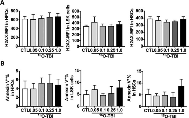
Fig 4. No changes were detected in DNA damage and apoptosis in HPCs, LSK cells and HSCs at three months after 16O TBI.
(A) Lin- cells were stained with an anti-γH2AX antibody and analyzed by flow cytometry. Data are presented as mean fluorescence intensity (MFI). (B) Isolated Lin- cells were stained with Annexin V to determine cellular apoptosis. Percentages of Annexin V positive cells are presented as means ± SD (n = 5).

