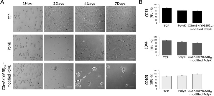
Fig 3. Effects of distinct substrates on hMSC morphology and surface marker regulation at passage 2.
(A) Phase contrast microscopy of adhering hMSCs onto TCP, PolyK and CGen3K(YIGSR)16-modified PolyK (scale bar = 150 μm), (B) expression of the multipotency markers CD73, CD44 and CD105. (Mean ± SD; n = 9).

