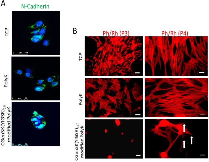
Fig 7. Expression and localization of N-cadherin and changes in cytoskeleton organisation within cells.
(A) Distribution of N-Cadherin (green staining) in relation to cell nuclei (blue) at P3 (scale bar = 50 μm), (B) cytoskeleton organisation on the different substrates. AT P4, hMSCs adhering on CGen3K(YIGSR)16-modified PolyK showed a tendency to form a cytoskeleton consisting of immature filaments (white arrows). (Scale bar = 50 μm).

