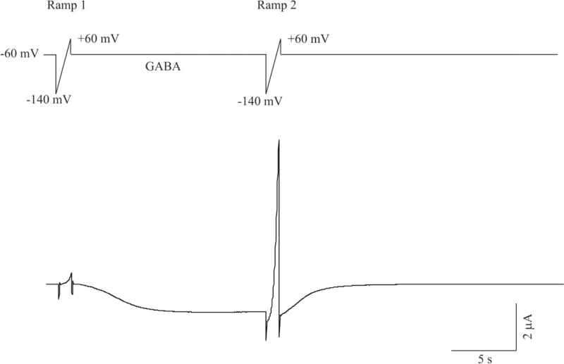
Figure 1.

Current recording protocols used for two-electrode voltage clamp recordings in Xenopus oocytes. Top: A given oocyte is voltage clamped at a holding potential of −60 mV and is activated first by a voltage ramp (Ramp 1, 1 sec duration, voltage changes at a rate of 1mV/5 ms from −140 mV to 60 mV), followed by a 15 sec pulse application of 1 μM GABA. Immediately following GABA application, a second ramp (Ramp 2) with identical features as the first one is applied. Bottom: A representative current trace evoked by this protocol in an oocyte expressing recombinant α1β2γ2S GABAA receptors.
