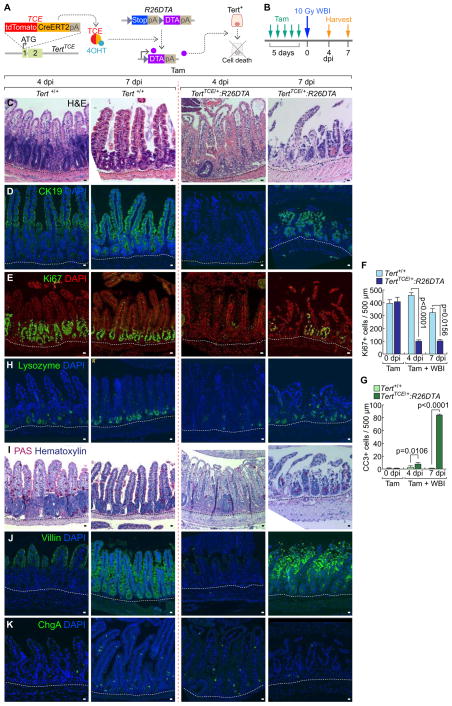
Figure 3. Impaired intestinal regeneration by conditional ablation of Tert+ cells.
(A) Illustration of conditional ablation of Tert+ cells (TertTCE/+:R26DTA). 4OHT treatment activates TCE, which leads to the expression of diphtheria toxin A (DTA) for Tert+ cell ablation.
(B) Scheme of mouse treatment.
(C–K) Impairment of intestinal regeneration by Tert+ cell ablation. H&E staining (C); cytokeratin 19 (CK19) (D); Ki67 (E); lysozyme (Paneth cell, H); PAS (goblet cell, I); villin (enterocyte, J); chromogranin A (ChgA; enteroendocrine cell, K). Of note, Tam-treated TertTCE/+:R26DTA did not affect intestinal homeostasis (Figures S3B–S3I). Quantification of the number of Ki67+ cells (F) and CC3+ cells (G) per 500 μm region of crypts. Scale bars=20μm; dot lines indicate the basal membranes below crypts.
The representative images are shown; N≥3.

