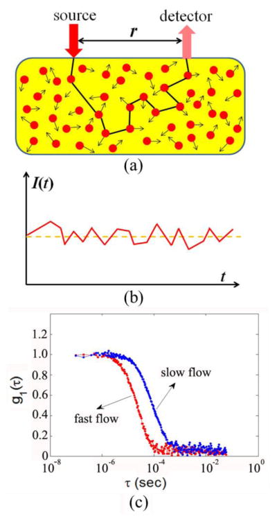
Fig. 1.
The principle of diffuse correlation spectroscopy (DCS) for blood flow measurements in deep/thick tissues. (a) Source and detector fibers are placed on the tissue surface within a distance of r for light delivery and collection; (b) Light intensity I(t), detected by the APD, fluctuates with time due to the motions of RBCs; (c) Blood flow change can be quantified from the shift of temporal electric field autocorrelation function curves.

