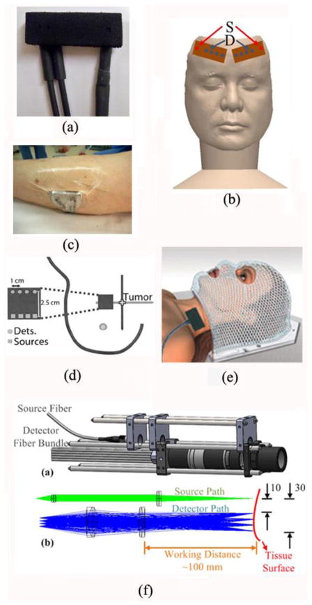
Fig. 3.
A variety of optical probes for DCS/DCT measurements. (a) A typical DCS probe consisting of source and detector fibers; (b) Two DCS probes taped on both sides of a frontal head for cerebral blood flow measurements; (c) A DCS probe taped on top of the calf muscle; (d) A hand-held DCS probe scanning over a breast tumor; (e) A DCS probe placed on top of the head/neck tumor; (f) A noncontact DCS probe with lens system focusing the source and detectors on the surface of tissue. Some of the figures are reproduced from the subfigures in the references (Huang et al., 2015c; Durduran et al., 2005)

