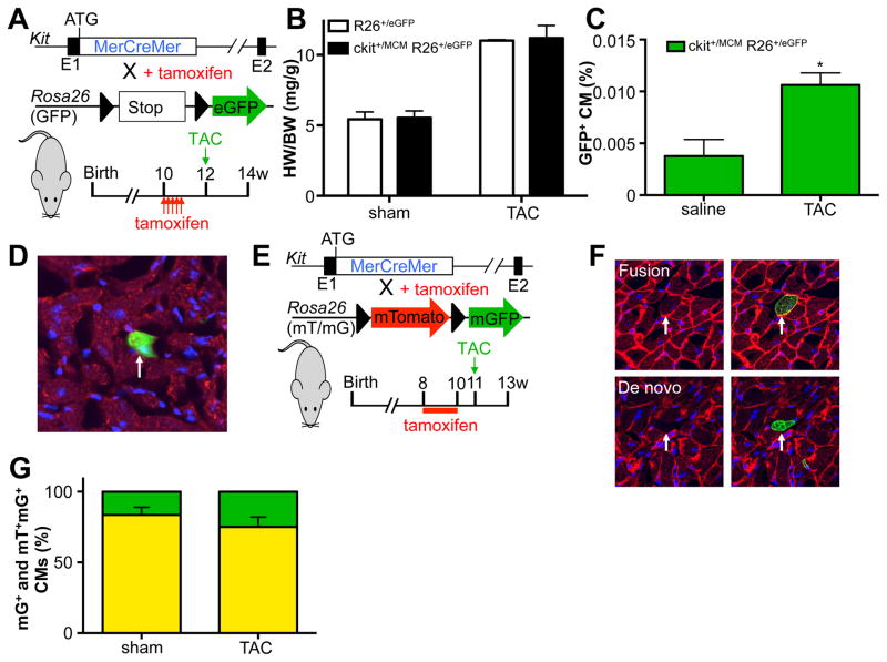
Figure 2. Pressure overload induced mild increase in c-kit derived cardiomyocytes.
A. Genetic model and experimental design. B. Pressure overload induces similar levels of cardiac hypertrophy in control and c-kit lineage tracing mice. C. Percentage GFP+ cardiomyocytes in response to sham or pressure overload (TAC) surgery. N=3 and 3 *p<0.05 D. Immunohistochemical staining of cardiac sections with DAPI (blue) and Desmin (red). Arrow indicates GFP+ cardiomyocyte. E. Genetic model and experimental design for panel F and G. F. Examples of fusion and de novo cardiomyocytes. G. Quantification of fraction of mGFP only (green) vs mGFP-mTomato dual positive (yellow) cardiomyocytes in response to sham or TAC surgery. N=7 and 6.

