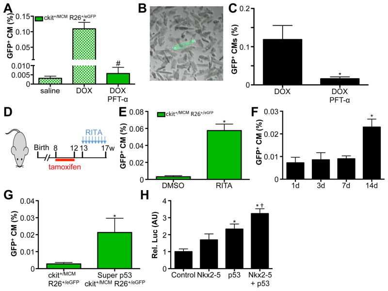
Figure 7. p53 mediates c-kit+ CPC derived cardiomyocyte formation.
A. GFP+ cardiomyocytes are completely blocked by injection of Pifithrin-α in conjunction with DOX. Blocked bars represent the same data as presented in Figure 4B. B. Adult cardiomyocyte isolation showing representative GFP+ adult cardiomyocyte. C. Quantification of isolated GFP+ adult cardiomyocytes shows pifithrin-α reduces abundance of GFP+ cardiomyocytes. N=4 and 3, *p<0.05 D. Experimental design used in panel E. E. RITA injection is sufficient to induce GFP+ cardiomyocytes. N=4 and 6, *p<0.05 F. Quantification of percentage GFP+ CMs in response to RITA administration. Hearts of mice were harvested at indicated time points after administration. N=3, 4, 4 and 4, *p<0.05. G. Quantification of percentage GFP+ CMs from Kit+/MCM X R-GFP and Super p53 Kit+/MCM X R-GFP mice. N=5 and 3, *p<0.05. H. Relative luciferase activity in HEK cells transfection with ANF promoter luciferase construct with control plasmid, 1μg Nkx2–5 expression plasmid, 1μg p53 expression plasmid and 0.5μg of both Nkx2–5 and p53 expression plasmid. N=3 each, *p<0.05 vs control, † p<0.05 vs Nkx2–5 and p53 alone.

