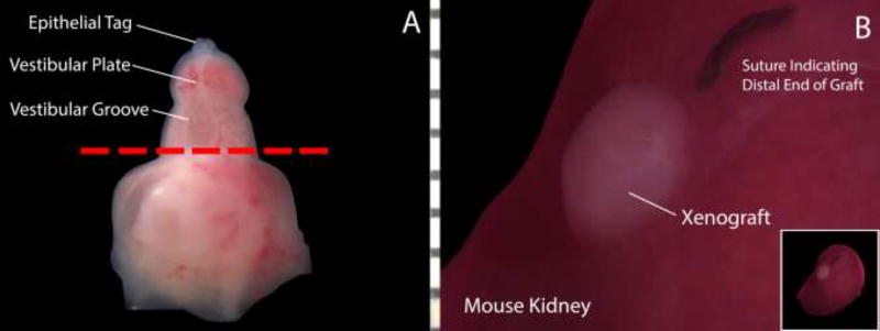
Figure 2.
Schematic demonstrating the xenografting process in a 10-week fetal clitoris. A: The distal genital tubercle is transected at the most proximal aspect of the open vestibular groove (red line) to ensure the absence of preexisting tubular structures and then grafted under the kidney capsule of an athymic mouse. B: Xenograft under mouse kidney capsule immediately prior to graft harvest. The nylon suture indicates the distal end/glans clitoris.

