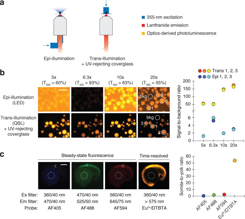
Figure 4. QSL trans-reflected illumination overcomes optics-derived photoluminescence.
(a) Optical paths of conventional epi-illumination (left) and trans-illumination (right) microscopy. The trans-illumination configuration also includes a UV-rejecting, TiO2-coated coverglass placed between the sample and the objective. (b) Eu3+/ATBTA-functionalized beads imaged by time-resolved microscopy, using the designated objectives and either LED epi-illumination or QSL trans-reflected illumination. Signal-to-background ratios for selected beads (dotted circles) are shown. Scale bar: 200 µm. (c) Zebrafish embryos immunostained with an anti-MYH1E primary antibody and a secondary antibody conjugated with the designated probe. Steady-state fluorescence and time-resolved lanthanide luminescence micrographs of 16-hpf embryos and their corresponding somite-to-yolk pixel intensity ratios are shown. Scale bar: 200 µm.

