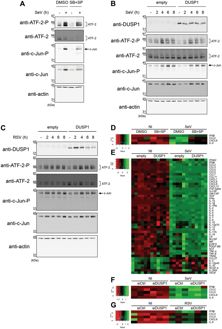
Figure 6.
DUSP1-mediated inhibition of JNK and p38 leaves virus-mediated activation of AP-1 and cytokine production intact. A549 cells were either pretreated with DMSO (vehicle) or SB203580 (10 μM) + SP600125 (10 μM) for 30 min prior to infection (A,D), transfected with empty or DUSP1-expressing plasmids (B,C,E) or transfected with Control (siCtrl) or DUSP1-specific siRNA (F,G) before infection with SeV at 40 HAU/106 cells (A,B,D,E,F) or RSV at MOI of 3 (C,G) for the indicated times or for 6 h (A,D), 16 h (E) or 12 h (F,G). In (A–C), levels of phosphorylated ATF-2 (ATF-2-P), total ATF-2, phosphorylated c-Jun (c-Jun-P), total c-Jun and DUSP1 were analyzed by immunoblot. Actin was used to verify equal loading. The data are representative of three independent experiments. Samples that are compared derive from the same experiment and blots were processed in parallel. Full-length blots are presented in Supplementary Figure 8. In (D–G), release of cytokines was quantified using Luminex-based multiplex assays. Heatmaps represent cytokine levels (pg/ml) logarithmically transformed, centered and scaled, measured in each biological replicates (n = 3 in D,F and G, n = 6 in E). Scatter plots of cytokine levels are shown in Supplemental Figure 1.

