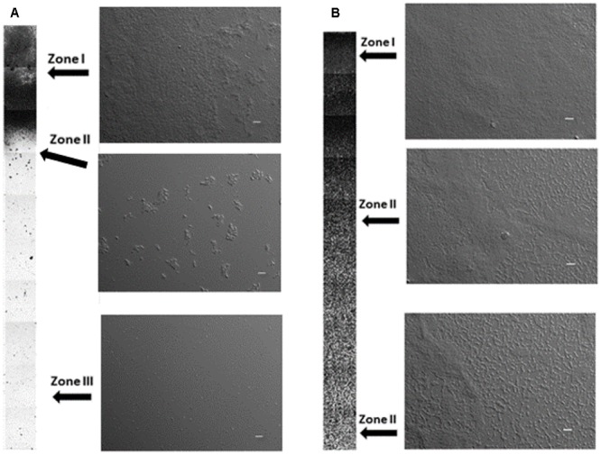
FIGURE 1.

Biofilms grown on glass surface in the rich medium supplemented with glucose (A), biofilms grown on PMMA surface in the rich medium supplemented with lactose (B) after 48 h of incubation. Zone I – arbitrarily designed high-density region where biofilm covered 90% or more of the available surface, Zone II – biofilm covered between 10 and 90% of the available surface, Zone III – biofilm surface coverage was less than 10%. Columns represent a low magnification DIC micrographs taken every 0.8 mm in the vertical direction from the water–air interphase (total depth 20 mm). Three representative higher magnification micrographs for different zones are shown on the right of the columns. Scale bar on micrographs represents 20 μm.
